
Echocardiography
Looking for an echocardiogram? At the WELL Health Clinic Network, we provide quick and convenient access to the care you need. Our dedicated healthcare providers are committed to delivering exceptional care, assisting you every step of the way.
Echocardiography Near You


Your Echocardiography Questions Answered
What is an echocardiogram?
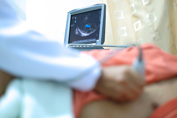
An echocardiogram (ECHO) is a non-invasive test that uses ultrasound waves to produce images of your heart.
Echocardiograms help your doctor visualize your heart’s structure, including the valves and chambers, and assess its function. Your doctor can use the images from an echocardiogram to identify heart disease.
When are echocardiograms used and what are the different types?
Your doctor may recommend an echocardiogram if they suspect issues with your heart’s valves or chambers or if you experience symptoms like shortness of breath or chest pain.
An echocardiogram can also detect congenital heart defects in unborn babies.
Depending on the information your doctor needs, you may undergo one of the following procedures:
- Transthoracic Echocardiogram: This is a standard, non-invasive echocardiogram. A technician (sonographer) will apply gel to your chest and use a device called a transducer to transmit ultrasound waves through to your heart. The transducer captures the echoes of the sound waves, converting them into moving images on a monitor. If your lungs or ribs block the view, a small amount of liquid (contrast agent) may be injected through an IV to enhance the visualization of your heart’s structure on the monitor.
- Transesophageal Echocardiogram: If getting a clear picture of your heart is difficult with a standard echocardiogram or if a more detailed visualization of the heart and valves is needed, your doctor may suggest a transesophageal echocardiogram. This test involves gently guiding a flexible tube with a transducer down your throat into the esophagus, which connects your mouth to your stomach. The transducer can then capture more detailed images of your heart.
- Doppler Echocardiogram: Sound waves bouncing off blood cells as they move through your heart and blood vessels produce Doppler signals, which help measure the speed and direction of blood flow. Doppler techniques are commonly used in transthoracic and transesophageal echocardiograms to detect blood flow problems and pressures in your heart’s arteries that traditional ultrasound may not identify.
- Stress Echocardiogram (Exercise Heart Evaluation): Some heart problems, particularly those related to the coronary arteries that supply blood to the heart muscle, only occur during physical activity. In a stress echocardiogram, ultrasound images of your heart are taken before and immediately after walking on a treadmill or riding a stationary bike. If you cannot exercise, a medication injection can simulate the effects of physical exertion, making your heart pump as if you were exercising.
How long does an echocardiogram take?
Most echocardiograms take less than an hour, but the timing may vary depending on your condition.
How do I prepare for an echocardiogram and what should I expect?
Before your echocardiogram:
- For a standard transthoracic echocardiogram, you don’t need to do any special preparation. You can eat, drink, and take medications as usual.
- If you have a scheduled transesophageal or stress echocardiogram, your doctor may instruct you to refrain from eating for a few hours before the test. Inform your doctor if you have difficulty swallowing, as it may affect the decision to perform a transesophageal echocardiogram.
- For a stress echocardiogram on a treadmill, wear comfortable shoes.
- For a transesophageal echocardiogram, make sure you arrange a ride home, as you’ll likely receive sedating medication.
During your echocardiogram:
- The technician will attach electrodes to your body to detect and conduct the electrical currents of your heart.
- Gel is applied to your chest to enhance sound wave conduction and eliminate air between your skin and the transducer.
- The technician will move the transducer over your chest to create recorded images on a monitor for your doctor to review.
- During a transesophageal echocardiogram, your throat will be numbed with a spray or gel to make inserting the transducer into your esophagus more comfortable. You’ll likely receive a sedative to help you relax.
- The technician may request certain positions or specific breathing patterns to obtain optimal images. Firm pressure may be applied to your chest, which could cause temporary discomfort.
After your echocardiogram:
- You can generally resume your regular activities following an echocardiogram.
- If your echocardiogram is normal, then further testing may not be necessary. If the results are concerning, you may be referred to a cardiologist for more tests.
- Treatment options depend on the exam findings and your specific symptoms. You may need a repeat echocardiogram in several months or other diagnostic tests, such as a cardiac computerized tomography (CT) scan or coronary angiogram.
For more information about our echocardiography services in Ontario, visit myhealthcentre.ca
How do I find an echocardiogram near me?
You can use the WELL Clinics website to quickly find an echocardiography clinic near you. If you have a referral or requisition, select the button below to book an appointment.
If you don’t have a referral or requisition but would like to request an echocardiography appointment, book with your family doctor. Alternatively, you can use our clinic map to find a primary care clinic or telehealth service and request a referral.